
TOWARDS A D3G GUIDE FOR DIAGNOSING MOLAR HYPOMIN
As noted here, D3G aims to develop guidelines that are both clinically useful and research friendly. Collaborative input is sought from the practitioner community, so please let us know what you think as our "draft-consensus" ideas evolve.
The Basics
As a general rule, Molar Hypomin is quite straightforward to diagnose once you know the ropes. We think it's important to not only recognise the key features of a "pristine case", but also to understand how and why these characteristics may change over time. And to be proactively vigilant for this common problem, it helps to know the "red flags" that might crop up on initial presentation.
Clinical Alerts
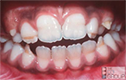
") There are three common features that may alert you to Molar Hypomin:
There are three common features that may alert you to Molar Hypomin:
- obvious Hypomin lesions ("demarcated opacities") – perhaps of aesthetic concern to the patient
- unusual decay – restricted to one or more molars, often contradicting good oral hygiene status
- dental pain – localised to the affected molars often
In any particular patient, the clinical appearance will depend to a large extent on how long the affected teeth have been erupted.
Spotting it Early
Generally a Hypomin Molar is easiest to spot soon after it emerges into the mouth – that is, before complicating factors set in. So for a typical case of Molar Hypomin – presenting either at 2 to 3 years (i.e. in "2-year", or "second baby" molars), or at 6 to 7 years (i.e. in "6-year" or "first adult" molars) – we like to diagnose at individual-tooth and whole-mouth (case) levels as follows:
At tooth level, a newly-erupted Hypomin Molar will typically exhibit:
- one or more "demarcated opacities", being spots or patches of opaque enamel with abnormal colour (white, cream, yellow or brown) and distinct boundaries with normal enamel
- such opacities have normal thickness of enamel throughout, but this may not last for long
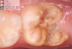
- likewise the opacity surface is initially intact, but this may not last for long as pictured here for a severely Hypomin 6-year molar undergoing "eruptive breakdown" while it's still erupting
- demarcated opacities are usually located in the cuspal half of the
crown and, where multiple opacities appear on a single tooth, they
may vary in colour, size and shape - Hypomin Molars may also elicit a painful response to mild stimuli
(tooth brushing, cold, heat, airstream)
At case level, a typical Molar Hypomin mouth may have:
- from one- to all four Hypomin molars of any type (obligatory)*, plus/minus...
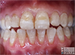
- one or more Hypomin adult incisors, generally less-affected than the molars (optional)
- *note the "sporadic presentation" of demarcated opacities (affecting different molars to different degrees) is a key diagnostic feature of Molar Hypomin
- also note that if only the incisors (but not the molars) have demarcated opacities then this isn't Molar Hypomin which by definition is "idiopathic" (i.e. the cause is unknown) – see "Local Hypomin" below
Spotting it Late
With time, a Hypomin Molar may change its initial appearance quite radically, reflecting inherent weaknesses of the abnormal enamel (i.e. soft, porous and prone to acid attack from caries & food/drink). And at whole-mouth (case) level, other clues and complications may manifest as follows:
Typically, severe Hypomin Molars will progress through these four steps:
- surface pitting of the demarcated opacity, perhaps associated with extrinsic staining
- further degradation and breakdown of the opacity, leaving sharp/fractured margins usually
- caries involvement (cavitation), with progressive loss of the Hypomin-enamel borders
- eventually, severe decay* may be all that remains apparent (see Unusual Decay pic above)
- *note we use the term "decay" to encompass physical breakdown (from chewing forces) plus acid attack from food/drink (enamel erosion) and dental plaque (caries)
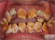
At later stages, a Molar Hypomin mouth may have:
- some molars with severe decay, and other teeth with Hypomin still visible
- unusual restorations in molars or incisors – location & shape more consistent with Hypomin (opacities) than with regular sites of plaque accumulation (gum line, deep occlusal fissures)
- one or more missing molars, extracted because of Hypomin (or the above sequelae)
Atypical cases of Molar Hypomin versus Local Hypomin
As noted above, a typical case of Molar Hypomin involves demarcated opacities on the 2-year molars ("Es") and/or 6-year molars ("6s"), with or without co-involvement of the adult incisors ("1s", "2s"). However, it's important to realise that other presentations can occur as any other tooth in the baby and adult dentitions can be affected by idiopathic demarcated opacities. When any molars (6s/7s/Es/Ds) are co-involved with teeth other than adult incisors, the case may be called atypical Molar Hypomin. Just to complicate things however, "non-idiopathic" demarcated opacities may also occur in other adult teeth (incisors, canines, premolars) as a result of localised disruption during enamel development – so-called "Turner teeth" (or incorrectly Turner's hypoplasia). Two common types of disruption are recognised, being trauma from, or infection of, the predecessor baby tooth. The term "Local Hypomin" is used in such situations of "ascribed causation", and the case can simply be described as "Local Hypomin affecting teeth X/Y/Z" – perhaps adding "secondary to [traumatised or infected] primary predecessor(s)".
The most common atypical Molar Hypomin cases include involvement of:
- baby canines ("Cs") and/or first molars ("Ds") – consistent with earlier causation
- adult canines ("3s") – particularly the cusp tips, consistent with later causation
- adult premolars ("4s" & "5s") or second molars ("7s") – consistent with even later causation still
The most common cases of Local Hypomin include involvement of:
- adult incisors only ("1s", "2s") – consistent with earlier disruption of corresponding baby incisors (e.g. traumatic injury or abcessed during infancy)
- premolars only ("4s, 5s") – consistent with earlier disruption (e.g. abscess and/or extraction) of corresponding baby molars during early childhood
- baby and adult teeth disrupted by surgical trauma (e.g. during cleft lip/palate surgery)
Checking it's not something else
To ensure your case is Molar Hypomin and not something else, a differential check can be run against the prime alternatives as follows:
1. Is it Amelogenesis Imperfecta?
|
|
Amelogenesis imperfecta |
Molar Hypomin |
|
Affected teeth |
All |
Molars, +/- adult incisors |
|
Family history |
Yes, almost always |
Not usually, but possible |
- see an example of AI here
2. Is it Dental Fluorosis?
|
|
Dental Fluorosis |
Molar Hypomin |
|
Affected teeth |
Many, mostly incisors |
Molars, +/- adult incisors |
|
Chronological pattern |
Symmetrical usually |
Often asymmetrical (sporadic) |
|
Lesion borders & opacity |
Diffuse borders, mottled |
Demarcated borders, dense |
|
Opacity colour |
White usually |
White or cream/yellow/brown |
- see an example of dental fluorosis here
3. Is it a Chronological Defect?
|
|
Chronological Defect |
Molar Hypomin |
|
Affected teeth |
Many, mostly incisors |
Molars, +/- adult incisors |
|
Chronological pattern |
Symmetrical |
Often asymmetrical (sporadic) |
|
Lesion feature |
Linear, right across tooth |
Non-linear, localised areas |
- see an example of chronological defect here
4. Is it Enamel Hypoplasia?
|
|
Enamel Hypoplasia |
Molar Hypomin (w. breakdown) |
|
Affected teeth |
Any, no bias towards molars |
Molars, +/- adult incisors |
|
Lesion border |
Rounded edge & regular outline |
Sharp edge & irregular outline |
|
Missing enamel |
Before, during & after eruption |
After eruption only |
- see an example of enamel hypoplasia here

5. Is it normal Dental Caries?
|
|
Dental Caries (normal enamel) |
Molar Hypomin (with caries) |
|
Affected teeth |
Any, not restricted to molars |
Restricted to molars often |
|
Usual locations |
Occlusal, interproximal, cervical |
Cuspal, occlusal – not cervical |
|
Plaque-associated |
Yes, often obviously |
Low-plaque areas often |
|
Hypomin enamel visible |
No |
Often yes (same or another tooth) |
- see an example of dental caries here (help: great images needed)